Figure 8.

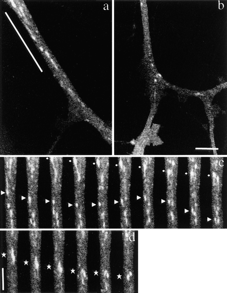
Axonal transport of vesicles containing GAP-43–GFP chimeric protein in mouse DRG neurons. Neurons were transfected with adenovirus vectors carrying GAP-43–GFP chimeric DNA after 3 h in culture and were observed using a laser scan microscope 40 h later. Note that numerous vesicles of various sizes were transported from the proximal side of the axon. (a and b) Proximal and distal area of the same axon. a shows an axon ∼40 μm from the cell body, b shows the same axon ∼110 μm from the cell body. The upper side of both micrographs is proximal to the cell body. Note the difference in the sizes of GAP-43–transporting vesicles even though the degree of magnification of both figures is the same. (c and d) Time course images of the underlined area of the axon in a. The intervals between frames are 3.45 s. The upper side of both micrographs is proximal to the cell body. In c, tubular and spherical vesicles of various sizes and shapes are moving. The vesicle indicated by diamonds showed representative continuous anterograde movement. The vesicle indicated by arrowheads switched back to retrograde movement, and again switched direction anterogradely. In d, the vesicle indicated by stars broke down into smaller spheres while moving anterogradely in the axon. Bars: (b and d) 5 μm.
