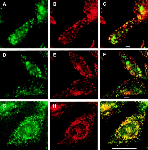
Figure 5.
Double-label immunofluorescence with internalized GLUT4 and transferrin. Cells were incubated with both Texas red–coupled transferrin and fluoresceinated 9E10 at 15°C for 2.5 h, and then either processed immediately for microscopy (A–F) or shifted to 37°C for 10 min followed by processing for microscopy (G–I). A, D, and G, FITC-9E10; B, E, and H: Texas red–transferrin; and C, F, and I: merged images. Areas of overlap are indicated in yellow. Bars: (A–F) 2 μm; (G–I) 10 μm.

