Figure 8.

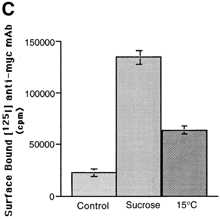
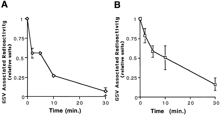
Redistribution of GLUT4 from GSVs to plasma membrane after inhibition of GSV formation. GSVs were labeled by incubating cells with radioiodinated anti-myc mAb for 80 min at 15°C, and then shifting cells to 37°C for 30 min. The rate of disappearance of GSVs was then assayed by incubating cells at 37°C in media containing 0.45 M sucrose (A) or at 15°C in regular media (B). GSV-associated radioactivity was determined for each time point and plotted as in Fig. 2 B. In parallel, the relative amount of GLUT4 on the cell surface under each of these conditions was also determined (C). Cells were incubated with hypertonic sucrose or incubated at 15°C in regular media for 30 min, fixed, and then incubated with radioiodinated 9E10 antibody for 1 h at room temperature, and washed extensively. Cell-associated radioactivity was then determined by counting cell pellets in a gamma counter. With the disappearance of GSVs in the presence of sucrose or with incubation at 15°C, there is a concurrent increase of GLUT4 on the cell surface.

