
Figure 2.
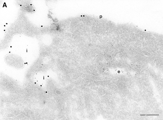
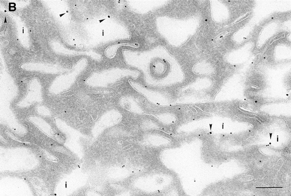
(A and B) Biotinylation of the surface of cells expressing ARF6(Q67L). Cells were either infected with vector virus and incubated with HRP for 10 min (A) or infected with recombinant virus encoding ARF6(Q67L) (B), and then incubated with sulfo-NHS-biotin and processed for immunolabeling. Sections were labeled first with an antibiotin antibody followed by protein A–gold (15 nm), and then with an antibody against HRP followed by protein A–gold (5 nm, A), or with antibody against ARF6 followed by protein A–gold (10 nm, B). (A) Only the plasma membrane and cross-sections across membrane invaginations were labeled with biotin. Early endocytic structures labeled with HRP were inaccessible to biotin. (B) In ARF6(Q67L)- expressing cells, plasma membrane invaginations were accessible to biotin (arrowheads) and thus are in continuity with the extracellular milieu. i, invaginations; p, plasma membrane; e, endosomes. Bar, 200 nm.