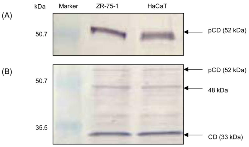
Fig. 1.

Analysis of pCD secretion by HaCaT and ZR-75-1 cells. HaCaT and ZR-75-1 cells were seeded in normal growth medium followed by replacing to serum-free media for additional 48 h. Conditioned media were collected and concentrated (10×). The cell pellets were lysed in lysis buffer. Protein contents of conditioned media and lysates were determined and equal amounts of total protein were analyzed by SDS-PAGE followed by immunoblotting. pCD protein in conditioned media was detected using anti-AP antibody (A); pCD and CD levels were detected in cell lysates using anti-CD antibody (B).
