Figure 2.
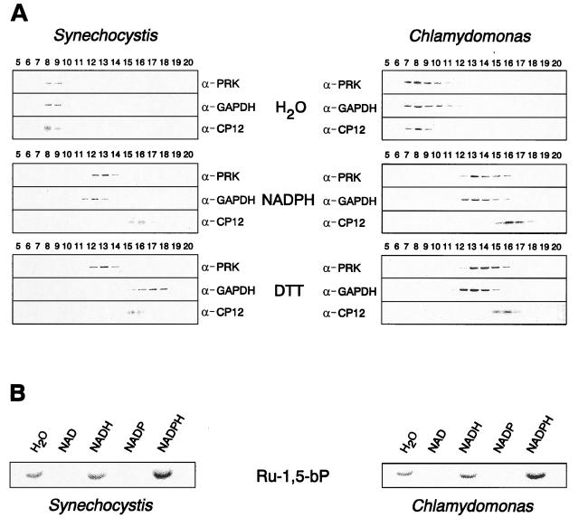
Identification and characterization of PRK/CP12/GAPDH complexes in Synechocystis and C. reinhardtii. (A) Isolated complexes remain stable, but dissociate in the presence of NADPH or DTT, respectively. Soluble supernatants of lysed Synechocystis and Chlamydomonas cells were subjected to size-exclusion chromatography. The pooled complex fractions 7–9, which contained the complex, were treated either with NADPH (2.5 mM, 30 min, 4°C) or with the reducing agent DTT (20 mM, 30 min, 30°C). For a control, they were incubated with the same volume of water (30 min, 4°C). All assay mixtures were rechromatographed on the same size-exclusion column, and the collected fractions were determined by immunoblot analysis using antisera (α) to PRK, GAPDH, and CP12. Lanes are numbered according to the collected column fractions. (B) PRK activity depends on dissociation of the PRK/CP12/GAPDH complex and NADPH. Pooled complex fractions 7 and 8 from size-exclusion chromatography (see above) were assayed for ribulose 5-phosphate phosphorylation activity dependent on the indicated nicotinamide-adenine dinucleotides (2.5 mM each). Formation of radiolabeled ribulose 1,5-bisphosphate (Ru-1,5-bP) was visualized after thin-layer chromatography and exposure of the plates to x-ray films.

