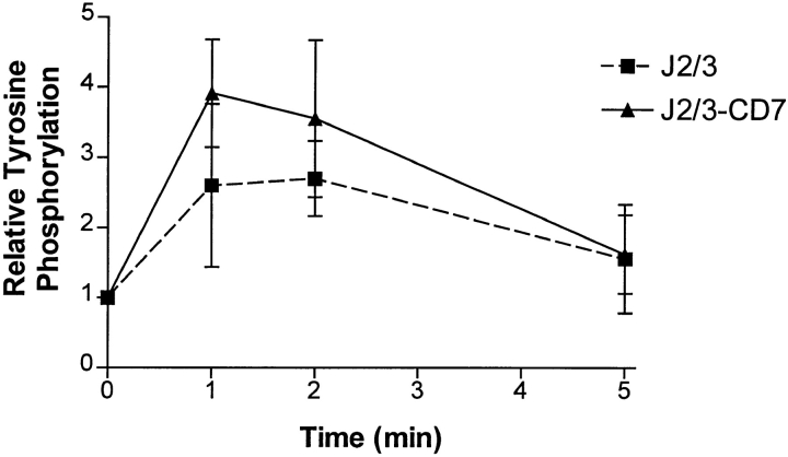
Figure 6.
The tyrosine phosphorylation of PLC-γ1 after crosslinking various FcγR. J2/3 (squares) or J2/3-CD7 (triangles) cells were incubated with mAbs IV.3 (anti-FcγRII) and 3G8 (anti- FcγRIII), warmed to 37°C, and crosslinking initiated by addition of F(ab′)2 fragments of goat anti–mouse antibodies. At each time point, an aliquot was removed, PLC-γ1 was immunoprecipitated, and proteins were separated by SDS-PAGE. Blots were probed with anti-phosphotyrosine and subsequently with anti–PLC-γ1 antibodies to determine the relative phosphorylation of the immunoprecipitated enzyme, as described in Materials and Methods. Three independent experiments from both cell types were analyzed by densitometry, and the mean and SEM of the three experiments are shown.

