
Figure 10.
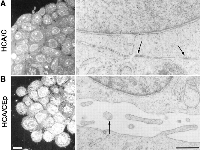
Ultrastructure of the aggregates formed by HCA/C and HCA/CEp cells. Reflection contrast micrographs of cross sections through the aggregates formed in 90 min by each cell type reveals a tight organization of the HCA/C aggregates and loosely interconnected cells forming the HCA/CEp aggregates. (A) Electron microscopy on the preparations shows the abundant presence of the adherens junctions between HCA/C cells (arrows). In contrast, microvilli (B, arrow) were found at the intercellular space between the cells of internal layers of HCA/CEp cells. In HCA/C cells, microvilli were present almost exclusively at the apical membrane of the outer layer of cells in the aggregate and not on the surface of the cells from internal layers. Bars: (left) 10 μm; (right) 0.25 μm.