
Figure 3.
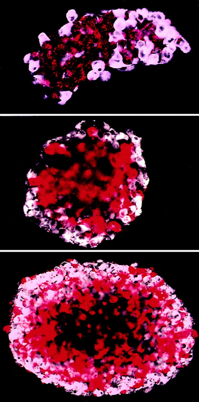
Segregation of Ep-CAM–positive LEC cell transfectants from the parental cells in multicellular aggregates. LEC-C and LEC-Ep cells, labeled with fluorescent dyes PKH-26 and PKH-2, respectively, were mixed at a 1:1 ratio, sedimented, and allowed to form an aggregate. This aggregate, in which both cell types were represented in a random pattern, was mechanically dispersed, and the smaller aggregates obtained were further cultured in suspension for 24 h, fixed, and analyzed. The micrographs present optical cross sections at the equatorial area of the aggregates after 24 h, as seen with a confocal microscope. The artificial colors were assigned to the cells depending on the color of the fluorochrome and the cell type: LEC-C (red); LEC-Ep (white) cells. The figures show similar cell patterning in different size aggregates in the range of <100 to ∼1,000 cells. A dark area in the middle of some aggregates is an optically nontransparent zone.