
Figure 3.
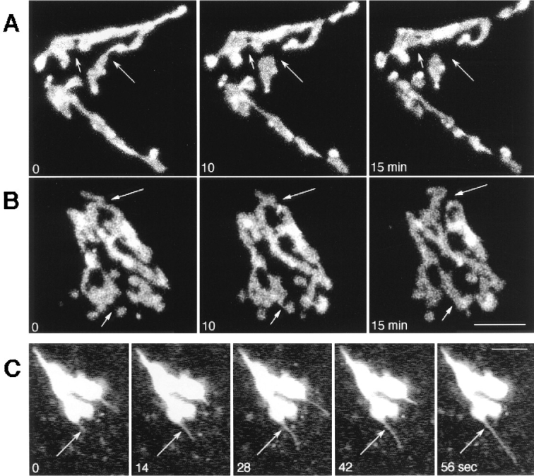
Tubule connections and extensions of the Golgi complex. (A and B) A time series of images from HeLa cells expressing GFP-GalTase (A) or GFP-KDELR (B) were collected at 0, 10, and 15 min at 37°C on a confocal microscope. Each image represents an overlay of a set of confocal slices extending the depth of the cell. Short arrows point to thin membrane connections initiating more stable membrane continuities between Golgi elements, while long arrows point to areas of detachment. (C) Single images with the confocal pinhole wide open were collected at 7-s time intervals in GFP-KDELR– expressing cells. Arrows point to thin tubule that rapidly extended off of Golgi rim. Bars, 3 μm.