
Figure 2.
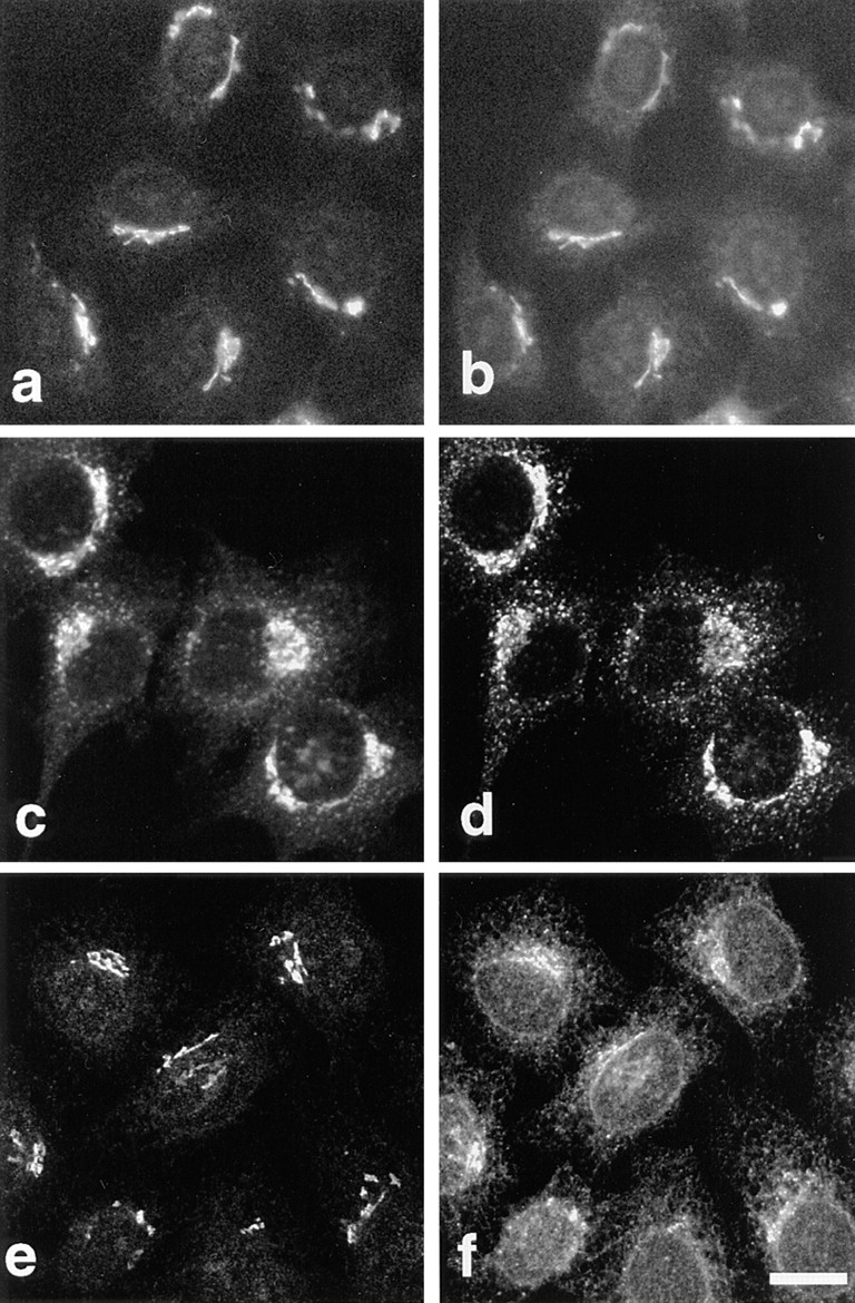
Immunolocalization of p23 to the perinuclear Golgi area. The p23 protein was decorated with antibody LP1 (a and e, HeLa) or CT (c, BHK). Double immunofluorescence with myc-tagged NAGT I (b), the KDEL receptor, ERD2 (d), and ERGIC- 53 (f) is shown. Bar, 5 μm.