
Figure 4.
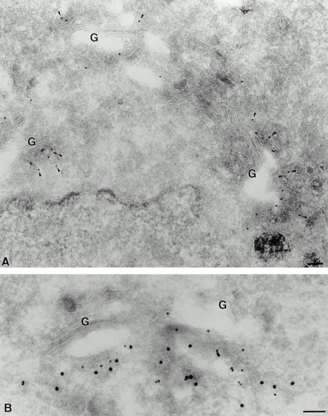
Localization of p23 to tubulovesicular membranes in the cis side of the Golgi. BHK cells were fixed and processed for frozen sectioning. A shows sections that were labeled with antibodies to p23 (CT) followed by 10 nm protein A–gold (arrowheads). Label is associated with tubulovesicular elements predominantly located in the vicinity of one face of the Golgi apparatus (G). Note the absence of labeling on other organelles including the ER surrounding the nucleus (lower left) and endosomes labeled with internalized 5 nm BSA-gold (lower right). B shows sections labeled with antibodies to p23 (CT; 15 nm gold) and to ERD2 (10 nm gold). The two antibodies colocalize on Golgi-associated membranes. Bar, 100 nm.