
Figure 6.
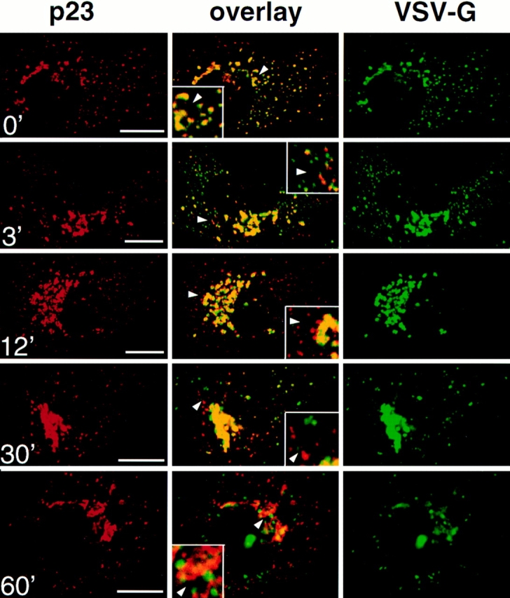
p23 colocalizes with VSV-G in the intermediate compartment at 15°C. Vero cells were infected with VSV tsO45 and maintained for 2.5 h at 39.5°C. Cycloheximide was then added, and tsO45-G was blocked in the intermediate compartment for 3 h at 15°C. The temperature was shifted to 31°C for the indicated periods of time, and cells were processed for double immunofluorescence with antibodies against p23 (CT) and VSV-G (P5D4). Images were processed and merged (overlay) as in Fig. 3. The inset shows a higher magnification of the area indicated by an arrowhead. VSV-G colocalized with p23 in the intermediate compartment at 15°C (0′). After release of the temperature block, forward transport of VSV-G resumed, and the degree of colocalization with p23 gradually decreased (3′, 12′, 30′, and 60′). Bar, 5 μm.