
Figure 5.
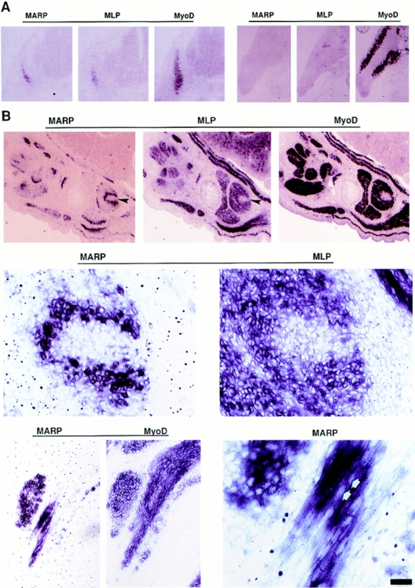
During muscle formation in vivo, appearance of MARP transcript coincides with terminal differentiation, and the transcript selectively accumulates at muscle end regions. In situ hybridization of mouse embryo sections. MyoD is expressed in myogenic cells before and after terminal differentiation; MLP is a marker for terminally differentiated striated muscle cells. The horizontal lines indicate consecutive sections (14 μm). (A) At E11.5, MARP mRNA is detectable within MLP-positive myotome (left), whereas in the forelimb, MyoD-positive myogenic progenitor cells were negative for MLP and MARP transcripts. (B) At E14.5, MARP transcript accumulated at end regions of MLP- and MyoD-positive muscles (top; the example is from foot muscles). A detail from the top panels (arrows) is shown in the middle panels. Note restriction of MARP transcript to edge of the indentation in the MLP-positive muscle. The bottom panels show an example of MARP transcript accumulation at the end of a MyoD-positive muscle (between ribs). In the corresponding higher magnification photograph on the right, MARP transcript can be detected inside striated cells, i.e., primary myotubes (arrows point to spared nuclei). Bar: (A) 480 μm; (B, top) 240 μm; (B, middle) 30 μm; (B, bottom, left) 60 μm; (B, bottom, right) 15 μm.