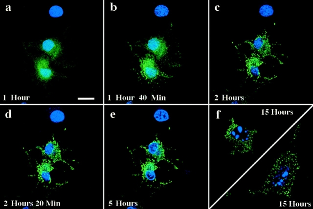
Figure 10.
GFP–Bax redistribution precedes nuclear fragmentation. A field containing three living Cos-7 cells expressing GFP–Bax was followed over time after addition of 1 μM STS, and 100 ng/ml of the nuclear stain bis-benzamide (a–e). In each panel, laser fluorescence confocal microscopy was used at the appropriate wavelength to visualize GFP (green) and bis-benzamide (blue). Time elapsed after STS addition is indicated in each panel. After 15 h (f) cells show fragmented nuclei associated with apoptosis. Bar, 20 μm.

