
Figure 9.
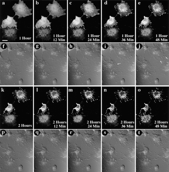
GFP–Bax redistribution occurs before cell shrinkage associated with apoptosis. A field containing three living Cos-7 cells expressing GFP–Bax was followed over time after addition of 1 μM STS. At each timepoint, the field was visualized by laser fluorescence to detect GFP–Bax (a–e and k–o), and by DIC to illustrate cell morphology (f–j and p–t). Time elapsed after addition of STS is indicated in the corner of each of the laser fluorescence panels. Redistribution of the GFP–Bax was first detectable for the two cells towards the top of the field at 1 h, 24 min after STS addition (c). Changes in cell shape first became apparent 12–24 min later, as evidenced by a retraction of cell outlines (arrows, i and j). Notice that at these timepoints, the lowermost cell has not yet initiated either GFP–Bax redistribution or cell shrinkage. GFP–Bax redistribution is first detectable for the lowermost cell at 2 h, 36 min (n). Bar, 20 μm.