
Fig. 4. Microtubules are required for grk RNA movement towards the ring canals and for transport into the oocyte.
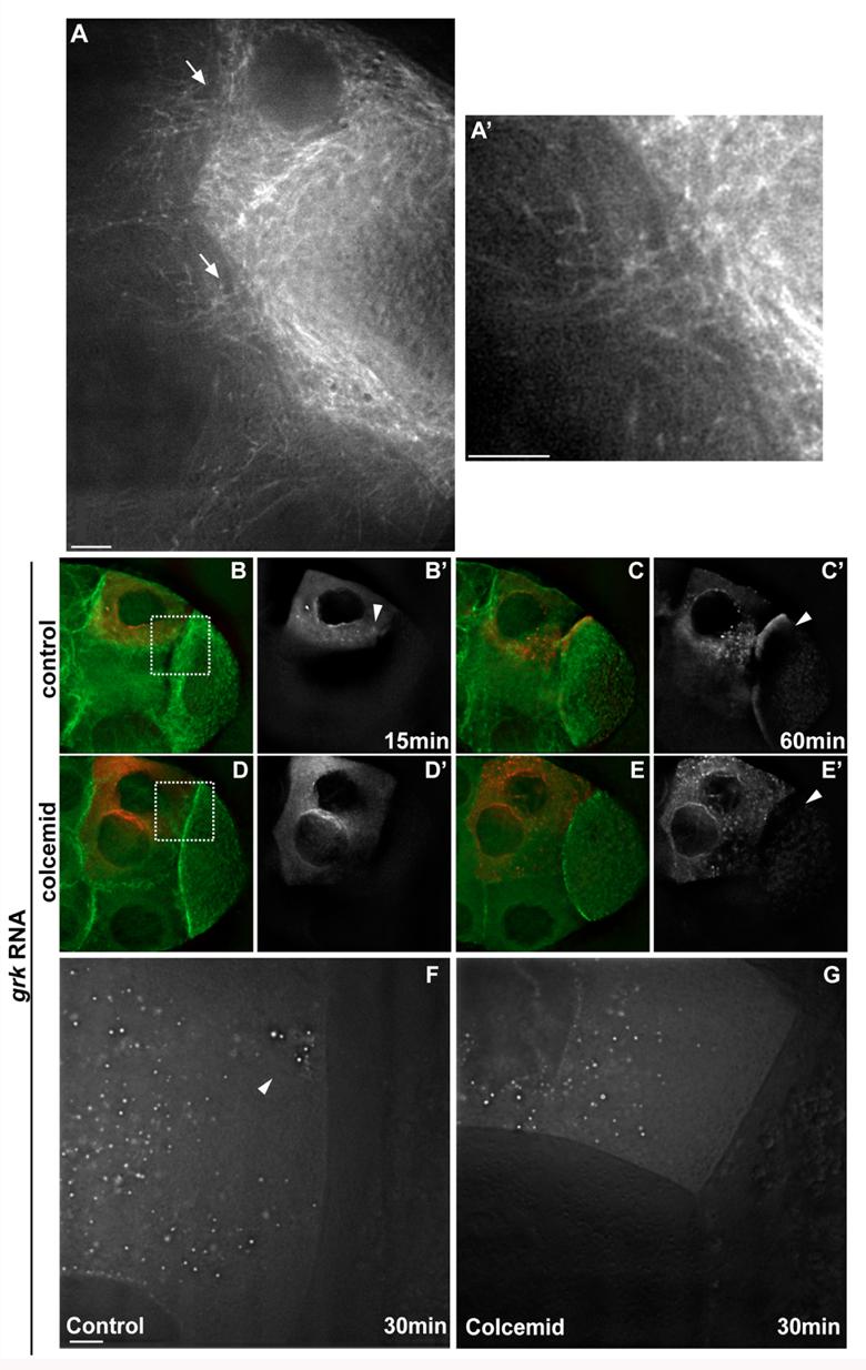
(A) Tau-GFP egg chambers show an enrichment of microtubules (MTs) at the ring canals (arrows). (A') Higher-magnification of ring canal in A. (B-E) Tau-GFP (green) living egg chambers injected with grk RNA (red) at 15 (B,D) and 60 (C,E) minutes after injection. (B,C) grk control; (D,E) grk-colcemid co-injection. (B',C',D',E') Only injected RNA is shown, at the corresponding time points. (F,G) Higher-magnification of boxed regions shown in B and D. Control (F) and Colcemid-treated (G) egg chambers at 30 minutes after injection with grk RNA. Notice how, in the absence of MTs, grk RNA particles fail to accumulate at the ring canals as they do in controls (F, arrowhead). In all panels, anterior is to the left and dorsal is to the top. Scale bar: 5 μm.