
Fig. 7. BicD protein is recruited by grk and bcd RNA and is necessary for grk RNA accumulation at ring canals.
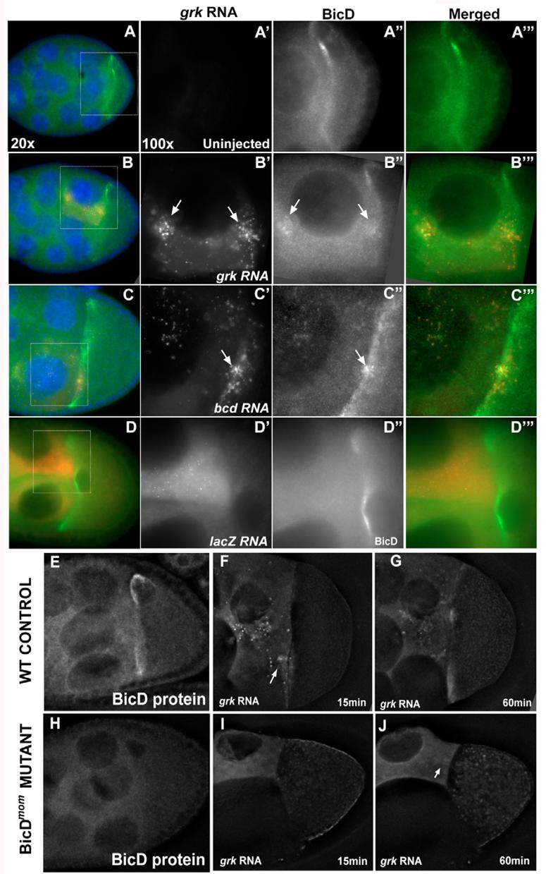
(A-D) Stage 8-9 egg chambers injected with RNA (red); BicD protein (green) and DNA (blue) are also shown (acquired at 20x). Outlined region represents the area shown (acquired at 100x) in the panels to the right (RNA, BicD and merged). (A-A‴) Uninjected egg chamber showing the BicD localization pattern around the oocyte nucleus. (B-C‴) Injected grk RNA (B′) and bcd RNA (C′) colocalize (arrows) with BicD (B″ and C″, respectively). (D-D‴) Injected lacZ RNA forms particles that do not accumulate (D′) and that do not colocalize with BicD (D″). (E,H) Immunofluorescence showing BicD localization in wild-type controls (E) and BicDmom (H) egg chambers. Notice the absence of BicD protein in BicDmom egg chambers (H). (F,G,I,J) Wild-type control (F,G) and BicDmom (I,J) egg chambers injected with grk RNA at 15 (F,I) and 60 (G,J) minutes after injection. In the absence (H-J) of BicD protein, grk RNA ring canal accumulation (arrow) and localization in the oocyte is strongly reduced. In all panels, anterior is to the left and dorsal is to the top (except D, dorsal side where the oocyte nucleus is).