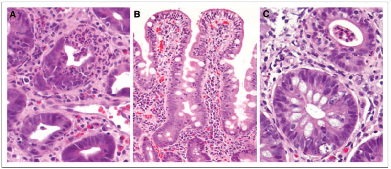
Fig 3.

Representative photomicrographs of histopathologic features of enterocolitis. (A) Neutrophilic infiltration with colonic crypt destruction (hematoxylin and eosin, ×400). (B) Small bowel mucosa showing markedly increased surface intraepithelial lymphocytes and expansion of lamina propria with mononuclear cells (hematoxylin and eosin, ×200). (C) Two adjacent colonic glands showing cryptitis in the upper gland and intraepithelial lymphocytosis and crypt cell apoptosis in the lower gland (hematoxylin and eosin, ×400).
