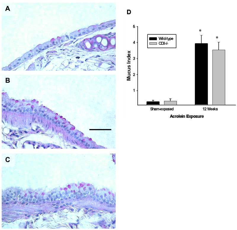
Figure 3. Epithelial cell hypertrophy and mucous cell metaplasia develop in mice exposed to acrolein for 12 weeks.
(A–C) Mucous cell metaplasia was visualized by light microscopy of periodic acid-Schiff-stained lung sections. Photomicrographs (400x original magnification) are representative of 8 mice per group. Scale bar = 50μm. (D) Mucous production by airway epithelial cells in acrolein-exposed mice is not different between wild-type and Cd8(−/−) mice. Mucous index was derived from n = 5 mice per group. Values presented are means ± sem. No significant differences were observed between wild-type and Cd8(−/−) mice exposed to acrolein for 12 weeks. * denotes value significantly greater than strain-matched sham-exposed control at p<0.05.

