
Figure 3.
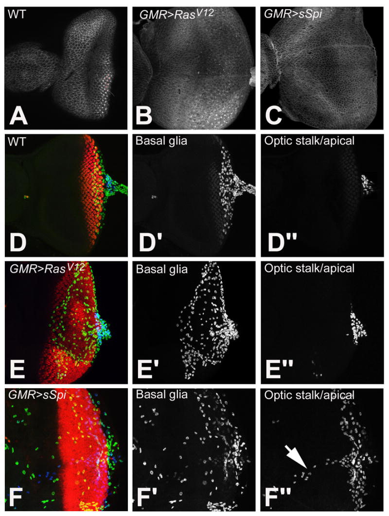
Examination of the PE in wild type (A), GMR>RasV12, (B) and GMR>sSpi (C) eye imaginal discs with Arm. Retinal basal glia in wild type (D-D″), GMR>RasV12 (E-E″) and GMR>sSpi (F-F″) eye imaginal discs. Differentiating photoreceptors are labeled with ElaV (red). Glia are identified with Repo. Glia in the basal part of the eye disc are in the green channel (D′, E′ and F′). Glia located apical and in the optic stalk are in the blue channel (D″, E″ and F″). Normally glia are located basal to photoreceptors and within the optic stalk (D′ and D″). This is unaffected in GMR>RasV12 (E′ and E″). In GMR>sSpi, basal glia migrate more anteriorly (F′), some glia are also located apically and along the Bolwig nerve (arrow) (F″).