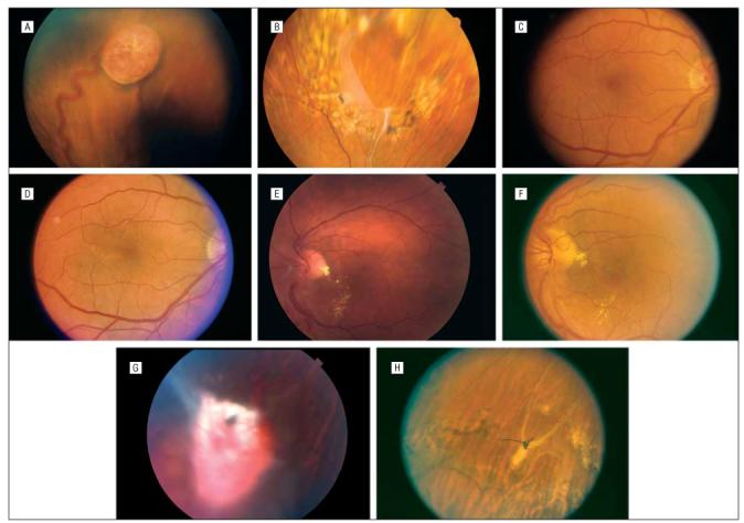
eFigure.

A-H, Views of patients 2 and 3. Patient 2, A-D, Before and after surgery. E, View of the resection site, with surrounding photocoagulation. Fibrosis hides the 10-0 polypropylene sutures. Patient 3, F-H. Before surgery and at 4 months after surgery, with partial resolution of macular exudates (F). Prominent fibrosis overlies the retinal hemangioblastoma (G). After surgery, the retina is attached via laser photocoagulation around the resection site (H).
