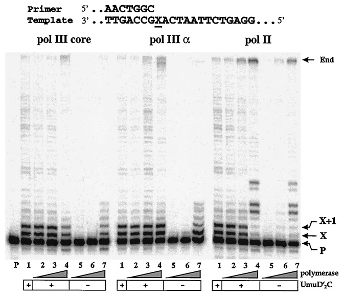
Figure 2.
Effect of pol III and pol II on translesion replication. Standard polymerization reactions, using a standing-start protocol, were carried out in either the presence or absence of UmuD′2C by using different concentrations of pol III core (0, 0.5, 2, 20 nM), pol III α-subunit (0, 0.5, 2, 20 nM), and pol II (0, 0.2, 1, 10 nM). All reactions contain RecA, β,γ-complex, SSB, four dNTPs (100 μM), and ATP (1 mM). Lane P contains the 32P-labeled primer in the absence of proteins. Locations of the unextended primer band, abasic site (X), downstream site adjacent to the lesion (X + 1), and end of template are indicated on the right. The DNA used in the standing-start protocol is shown at the top.

