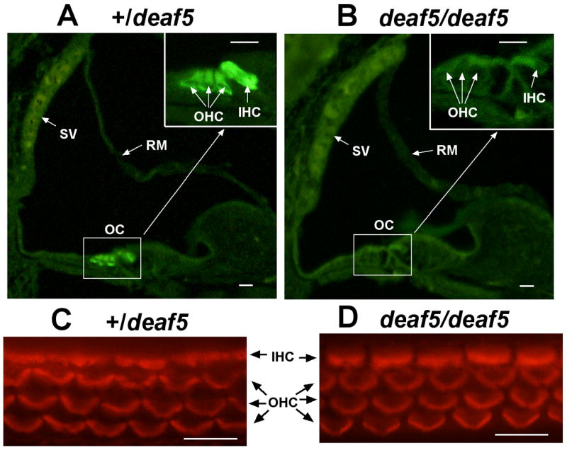
Fig. 3. Absence of OTOF in cochlear hair cells of deaf5/deaf5 mutant mice.

Cross sections through one turn of the cochlea from a P6 +/deaf5 control mouse (A) and a P6 deaf5/deaf5 mutant (B). The boxed region enclosing the organ of Corti (OC) is enlarged in the upper righthand corner in both A and B, and the stria vascularis (SV) and Reissner’s membrane (RM) are indicated by arrows. OTOF-specific immunofluorescence was present in inner hair cells (IHC) and outer hair cells (OHC) of the +/deaf5 control but was not detected in hair cells of the deaf5deaf5 mutant. Phalloidin staining of organ of Corti surface preparations from P6 +/deaf5 controls (C) and littermate deaf5/deaf5 mutants (D) verify that inner and outer hair cells are intact in mutant mice. All scale bars, 10 μm.
