
Figure 6.


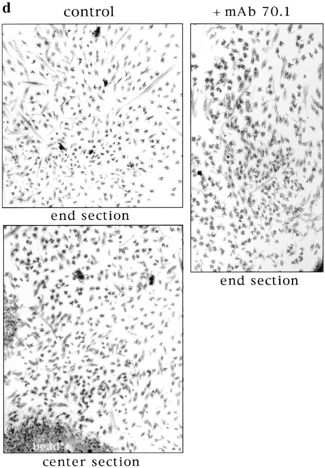
Microtubules are sorted into antiparallel arrays around chromatin in the absence of pole formation. (a) Immunofluorescent localization of NuMA to the frayed ends of chromatin bead spindles that have formed in the absence of dynein activity. (b and c) Hooking analysis. (b) Quantification of hook handedness in control and poleless spindles. Percentage of right- and left-handed hooks in sections through spindle centers containing chromatin beads, and spindle ends containing microtubule poles (control), or bundles (+ mAb 70.1). (c) Low magnification micrographs (5-μm width) are shown to give an overall impression of microtubule organization seen in sections through spindle ends containing microtubule poles or bundles and through spindle centers containing beads. (d) Higher magnification micrographs (2–2.5-μm width) show hooks on cross sectioned individual microtubules. In the presence of control antibodies, a section through a pole contains right-handed (clockwise) hooks, while a section containing beads contains both right- and left-handed hooks. In the presence of mAb 70.1, a section through a microtubule bundle likely to be at the spindle end is shown that contains almost exclusively left-handed hooks. Note: Hook handedness does not give any information about the polarity of the microtubules in these sections but indicates the degree to which microtubule polarity is uniform. 50–100 microtubules were evaluated in each section, and three sections were evaluated for each condition. Bar, 5 μm.