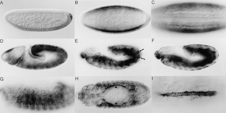
Figure 4.
Spatial expression pattern of mbc mRNA in wild-type embryos. In all panels, anterior is to the left. In A, D, E, F, and G, dorsal is at the top. (A) Lateral view, early stage 4, before cellularization. (B) Dorsal view, stage 5. (C) Ventral view, stage 6; the invaginating ventral furrow is evident. (D) Lateral view, stage 9. (E) Lateral view of the ectoderm, late stage 12; arrows highlight ectodermal stripes. (F) Lateral view focusing on the mesoderm and endoderm of the same embryo as in E. (G) Lateral view, stage 14; focusing on mesodermal cells. (H) Dorsal view, stage 14; arrowheads indicate the visceral musculature. (I) Dorsal view; stage 16; expression is evident in the cardial and pericardial cells of the dorsal vessel.

