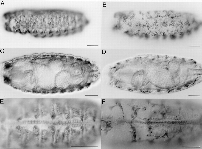
Figure 7.
Analysis of mesodermal derivatives in mbc mutant embryos. Tissues were visualized with a monoclonal antibody to MHC. All embryos are oriented with anterior to the left. A and B are lateral views with dorsal to the top, C and D are ventral views, and E and F are dorsal views. A, C, and E are wild-type embryos; B, D, and F are mbc F12.7/Df(3R)mbc-30 transheterozygotes. (A and B) Somatic muscle pattern of stage 16 embryos. Defects in myoblast fusion, as previously described by Rushton et al. (1995), are evident in B. (C and D) Visceral musculature and gut formation in late stage 16 embryos. Note the midgut constrictions in C and the absence of these constrictions in D. (E and F) Dorsal vessel of stage 17 embryos. At this level, there are no obvious defects. Bars, 50 μm.

