
Figure 8.
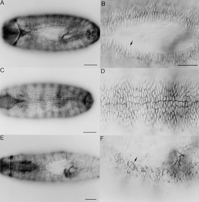
Defects in dorsal closure in mbc mutants revealed by staining with Fasciclin III. All panels are dorsal views with anterior to the left. A–D are wild-type embryos while E and F are mbc F12.7/ Df(3R)mbc-30 transheterozygotes. A and B show a stage 15 embryo in the process of dorsal closure. Arrow in B denotes elongated cells at the leading edge. C and D show a stage 16 embryo that has completed dorsal closure. E and F show a stage 16 embryo that has a pronounced defect in dorsal closure. Arrow denotes cells that are misshapen and have an improper accumulation of Fasciclin III along the leading edge. Bars: (A, C, and E) 50 μm; (B, D, and F) 25 μm.