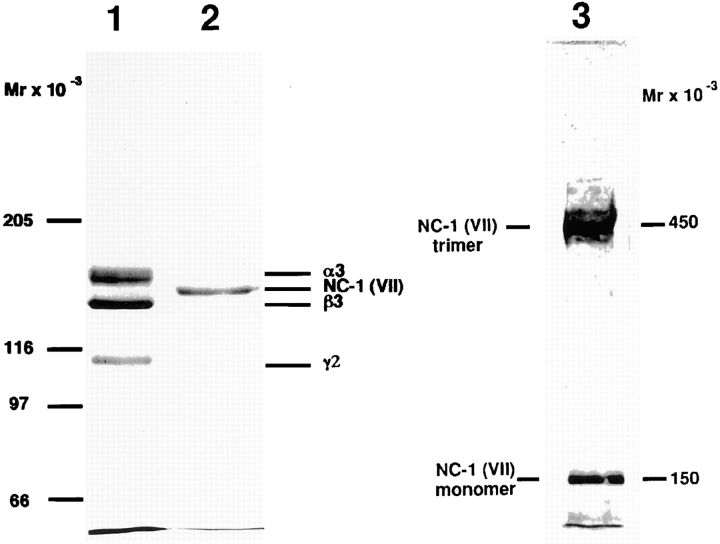
Figure 1.
Electrophoretic analysis of immunoaffinity-purified laminin 5 from cell culture media and type VII collagen–NC-1 from collagenase extracts of amniotic membranes. 2 μg of materials affinity purified on mAb 6/F12 from SCC25 cell culture medium (lane 1), and 2 μg of materials affinity purified on mAb NP-32 from collagenase extracts of amniotic membranes (lane 2) were separated by SDS-PAGE after reduction on a 3–7.5% gradient polyacrylamide gel and visualized by Coomassie blue staining. Consistent with previous results, the reduced bands representing subunits of laminin 5 (α3, 165 kD; β3, 140 kD; γ2, 155 and 105 kD) and type VII collagen–NC-1 (150 kD). The electrophoretic migration position of purified NC-1 before disulfide-bond reduction is shown in lane 3. Migration positions of the molecular weight markers are indicated to the left of each gel.

