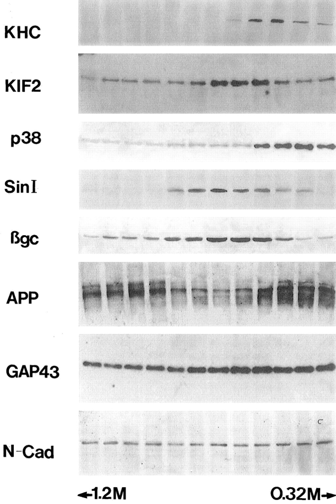
Figure 2.

The binding of KIF2 to membrane vesicles. Microsome fraction from developing rat cerebral cortex was fractionated by sucrose gradient centrifugation, and the same volume from each fraction was applied to SDS-PAGE, transferred to PVDF membranes, and analyzed by immunoblotting with antibodies against KHC, KIF2, synaptophysin (p38), synapsin I (Sin I), βgc, APP, GAP-43, and N-cadherin (N-Cad). Note that the peak fractions of KIF2 and βgc are highly coincident.
