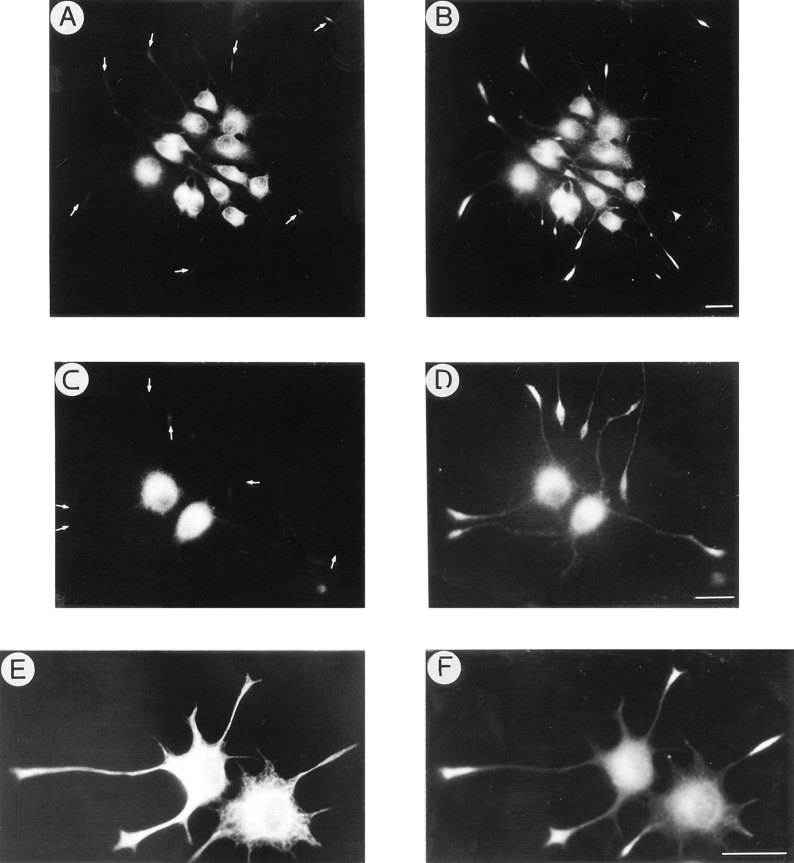
Figure 8.
KIF2 suppression alters the distribution of βgc, but not of synaptophysin, GAP-43, or synapsin I in NGF-treated PC12 cells. (A–D) Double immunofluorescence micrographs showing the distribution of βgc (A and C), synaptophysin (B), and GAP-43 (D) in NGF-differentiated PC12 cells treated with a KIF2 antisense oligonucleotide (ASKF2a, 5 μM). Note that while βgc completely disappears from growth cones (arrows), synaptophysin and GAP-43 remain highly concentrated at neuritic tips. (E and F) Double immunofluorescence micrographs showing the distribution of tyrosinated α-tubulin (E) and synapsin I (F) in NGF-differentiated PC12 cells treated with ASKF2a (5 μM). Note that synapsin I is highly concentrated at neuritic tips. For these experiments, cells were treated with oligonucleotides as described in Fig. 7. Bar, 10 μm.

