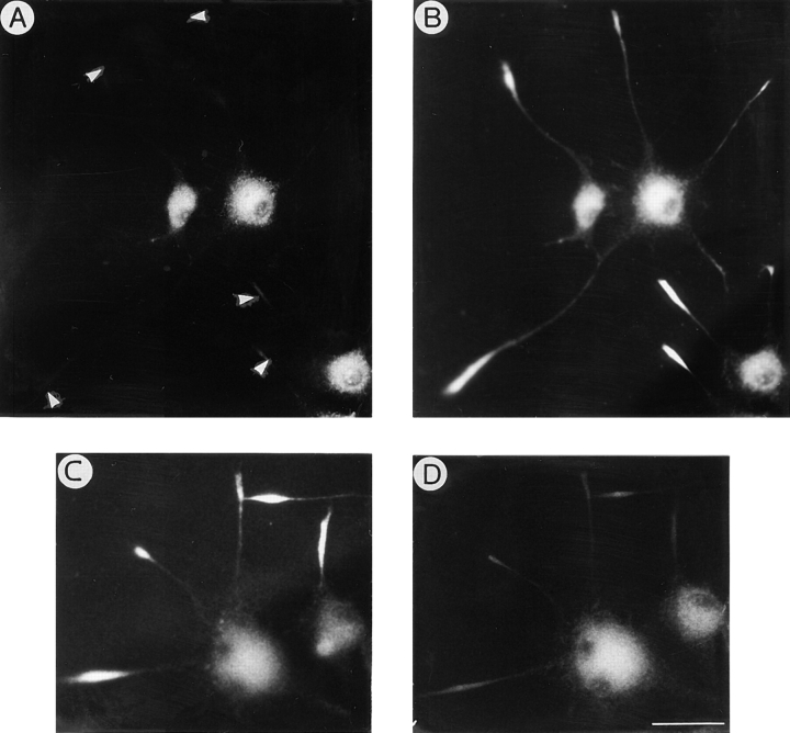
Figure 9.
(A and B) Double immunofluorescence micrographs showing the distribution of βgc (A) and APP (B) in NGF-differentiated PC12 cells treated with the KIF2 antisense oligonucleotide ASKF2b (5 μM). Note that while βgc completely disappears from growth cones (arrowheads) APP remains highly concentrated at neuritic tips. (C and D) Double immunofluorescence micrographs showing the distribution of βgc (C) and APP (D) in NGF-differentiated PC12 cells treated with the KHC antisense oligonucleotide -11/ 14 hkin (50 μM). Note that while this treatment does not affect the growth cone localization of βgc, it dramatically decreases APP immunofluorescence at neuritic tips. For these experiments, cells were treated with antisense oligonucleotides as described in Fig. 7. Bar, 10 μm.

