
Figure 6.
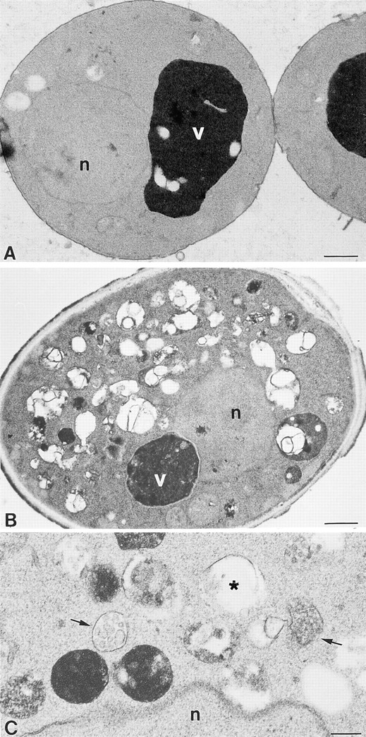
Ultrastructural analysis of vam3tsf mutants. (A) A cross-section of a typical vam3tsf (TDY1 + pVAM3-6.414) cell grown at 26°C, which closely resembles wild-type cells. (B) A cross-section of a vam3tsf cell after temperature shift to 38°C for 3 h. The accumulation of novel compartments is seen in addition to a prominent, electron-dense vacuolar compartment. (C) Enlarged examples of structures seen in vam3tsf cells grown at 38°C for 3 h. (Arrows) Multivesicular bodies; (asterisk) electron-transparent membrane compartment. n, nucleus; v, vacuole. Bars: (A and B) 500 nm; (C) 200 nm.