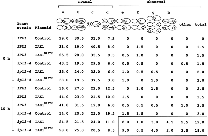
Figure 11.
Expression of IAK1 causes microtubule defects in ipl1 mutant yeast cells. Isogenic wild-type IPL1 (CCY98-3D-1) and mutant ipl1-4 (CCY98-3D-1-1) cells carrying the indicated plasmids (see Fig. 10 for description) were grown to early log phase at 30°C in supplemented minimal SD liquid medium (lacking uracil) that contained 2% raffinose as the sole carbon source (noninducing). At 0 h, galactose was added to give a final concentration of 4% (inducing), and the cultures were incubated at 30°C for another 10 h. At the time indicated, cells were fixed with formaldehyde, and the distribution of chromosomal DNA and microtubules in these cells were examined by indirect immunofluorescence microscopy. For each sample, 200 large-budded cells were scored, and the percentage of cells belonging to each cytological class is shown here. The classes represent: (a) cells with unseparated chromatin mass and a short to medium bipolar mitotic spindle; (b) cells with chromatin mass that is not fully separated and with an elongated, bipolar mitotic spindle; (c) cells with evenly separated chromatin masses and an elongated, bipolar mitotic spindle; (d) cells with evenly separated chromatin masses and a partially disassembled mitotic spindle or no mitotic spindle; (e) cells with unseparated chromatin mass, a single unduplicated or unseparated MTOC (spindle pole body), and an apparently monopolar spindle or no mitotic spindle; (f) cells with unseparated chromatin mass, a single unduplicated or unseparated MTOC, and no other microtubule structure; (g) cells with no MTOC or microtubule structure; and (h) cells clearly with unevenly separated chromatin masses. The boxed region highlights the abnormal phenotypes in ipl-4 yeast expressing the wild-type and IAK1D287N mammalian proteins.

