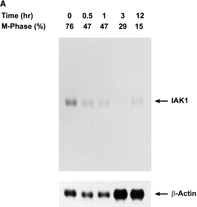
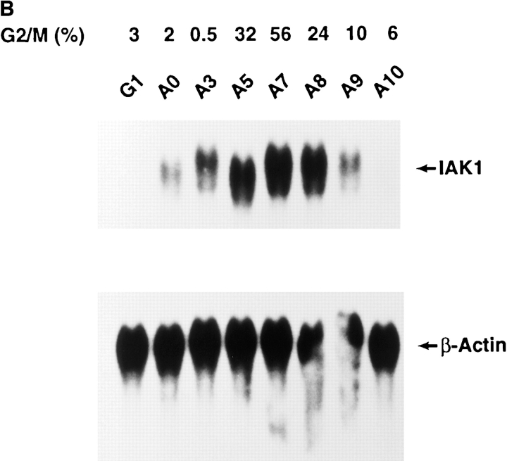
Figure 4.
Cell cycle analysis of IAK1 expression by Northern blot analysis. (A) Nocodazole block and release: Cells were serum-starved for 48 h in 0.5% β serum–containing medium. After resuming growth in a medium containing 10% serum for 12 h, cells were incubated in a medium containing 0.4 μg/ml of nocodazole for 18 h. Mitotic cells were collected by mechanical shake-off, replated into medium without nocodazole, and allowed to progress though the cell cycle. (B) Aphidicolin block and release: NIH 3T3 cells were serum-starved for 48 h in medium supplemented with 0.5% serum. The starved cells were released into the medium containing 10% serum and 5 μg/ml of aphidicolin for 18 h. After the aphidicolin block, the cells were washed three times in serum-free medium and released into medium containing 10% serum without aphidicolin. Cells were collected at different times after the release, and total RNA was isolated. Northern blot analysis of total RNA isolated from aphidicolin- and nocodazole-blocked and released cells were performed as described above except blots were reprobed with a β-actin probe to quantitate loading. The degree of synchrony of NIH 3T3 cells was determined by propidium iodide staining and flow cytometry and is shown as the percentage of cells of G2/M phase.


