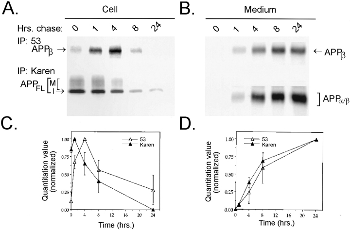
Figure 6.
Pulse–chase labeling demonstrates that intracellular APPβ is produced in an intracellular compartment before secretion in NT2N neurons. NT2N neurons were pulse labeled with [35S]methionine for 1 h and chased for 0, 1, 4, 8, and 24 h. Radiolabeled cell lysates (A) or media (B) were immunoprecipitated sequentially with antibody 53 (for APPβ) followed by Karen (for APPFL in the cell lysates and APPα/β in the media). Radiolabeled immunoprecipitates were used to expose PhosphorImager plates (72 h) or X-ray film (3 wk) for visualization. C and D summarize the quantitation of experiments shown in A and B. Counts from three different experiments were normalized to percentage of maximum and plotted as shown (mean ± standard error).

