
Figure 10.
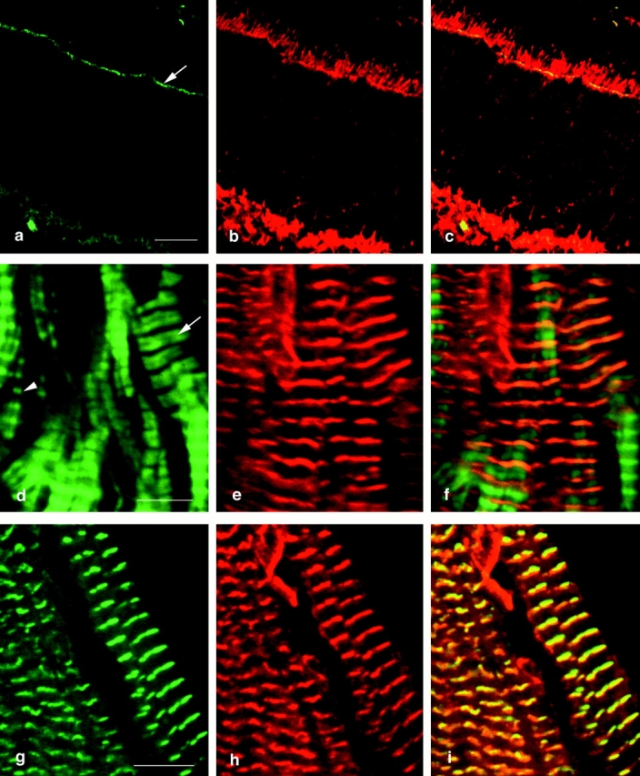
Colocalization of abLIM proteins with cytoskeletal proteins in adult murine retina and cardiac muscle. Murine retinal sections (a–c) were used to colocalize F-actin and abLIM. F-actin was visualized using BODIPY FL-conjugated phallacidin (a, green) and abLIM was visualized using specific primary antibodies followed by Cy3-conjugated secondary antibodies (b, red). Superimposed confocal microscope images identify coincident F-actin and abLIM staining in the actin-rich outer limiting membrane (c). F-actin (green) and abLIM (red) are similarly stained in unfixed sections of murine cardiac muscle (d–f). The periodicity of actin repeats varies across the section with areas of highly contracted muscle at left (arrowhead) and relatively relaxed muscle fibers at right (arrow). In the areas of the more relaxed fibers, abLIM is centered over the broad band of actin staining. Costaining of cardiac muscle with antibodies specific for mouse anti–α-actinin (g, green) and rabbit anti-abLIM (h, red) confirms the coincidence of the two proteins at the Z disks in cardiac muscle (i). Bars: (a–c) 20 μm; (d–f) 5.2 μm; (g–i) 5.6 μm.