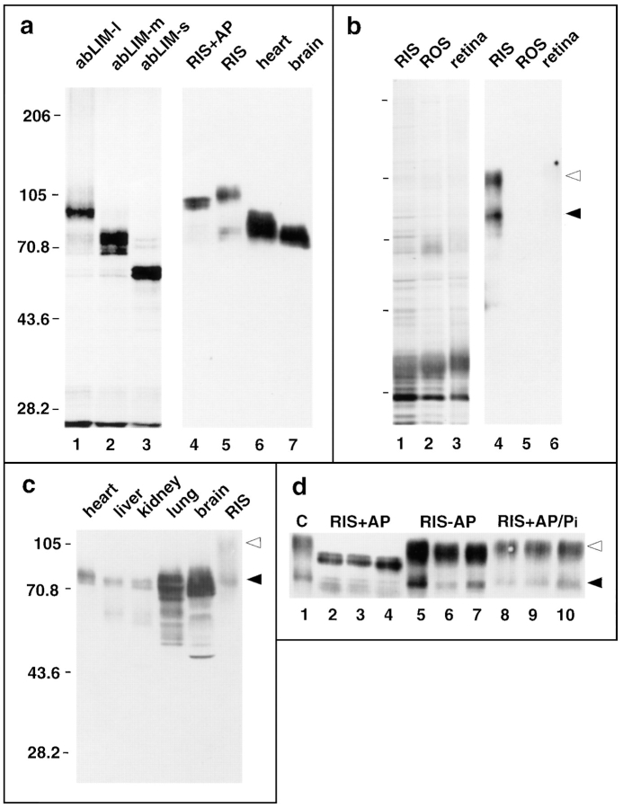
Figure 8.
Characterization of expressed abLIM polypeptides. (a) Comparison of abLIM polypeptides expressed in vitro and in vivo. 35S-labeled polypeptides expressed in vitro in reticulocyte lysates (a, lanes 1–3) are compared with abLIM polypeptides in immunoblots of extracts from various tissues (a, lanes 5–7). The additional bands below the major products in lanes 1–3 are probably due to internal initiation products from methionines 213, 324, 446, 582, and a cluster of nine methionines near the COOH terminus (between amino acids 696–771). Both sets of polypeptides were run on the same gel and prestained mobility standards were used to align the Western blot and the 35S-fluorogram. (b) A silver-stained SDS gel profile (lanes 1–3) and immunoblot (lanes 4–6) of retinal fractions. (c) Immunoblot of cytoskeletal extracts from various tissues. The SDS gel was overloaded to show minor polypeptide species. (d) Immunoblot of retinal extracts incubated for 15 min (lanes 2, 5, and 8), 30 min (lanes 3, 6, and 9), or 1 h (lanes 4, 7, and 10) with/without the inclusion of alkaline phosphatase. Tissue extracts in all panels are cytoskeletal fractions from heart, liver, kidney, lung, and brain. Mobility standards (kD) are indicated at left of each panel. (Filled arrowhead) abLIM-m; (open arrowhead) abLIM-l at ∼70 and ∼105 kD, respectively. abLIM-specific antiserum was used at 1:5,000. No staining was observed at 1:5,000 using preserum or serum absorbed with the GST–dem fusion protein (data not shown). RIS, rod inner segments; ROS, rod outer segments; retina, whole retina; C, control (untreated) rod inner segment extract; RIS+AP, RIS incubated with alkaline phosphatase; RIS–AP, RIS incubated without addition of alkaline phosphatase; RIS+AP/Pi, RIS treated with alkaline phosphatase in the presence of excess inorganic phosphate.

