
Figure 9.
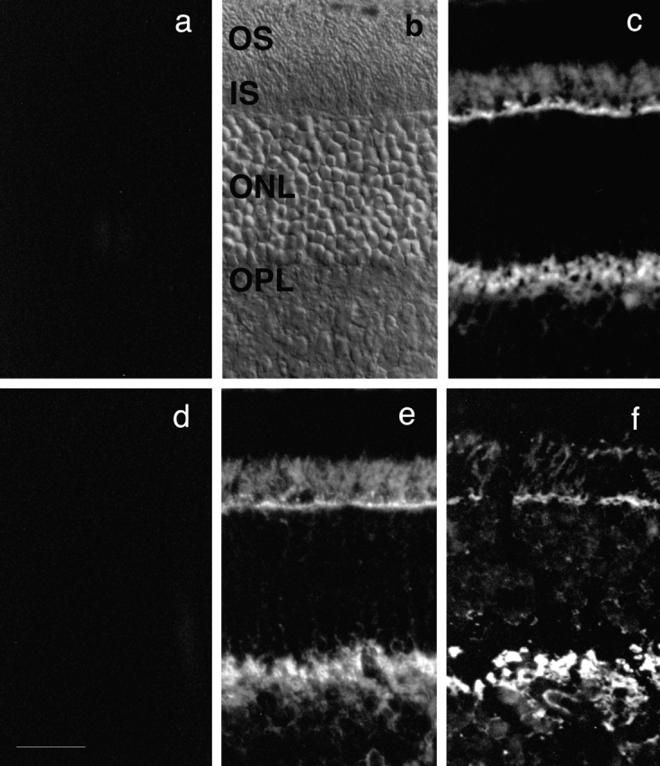
Localization of abLIM within retinal tissue sections. Unfixed cryostat sections prepared from adult murine retinas (a–e) or an adult human retina (f). Sections in c and f were treated with abLIM-specific antiserum at 1:500. Staining is prominent in the inner segment layer (IS) and outer plexiform layer (OPL). Staining is abolished in control sections treated with preimmune rabbit serum (a) or abLIM-specific serum preabsorbed with the GST–abLIM fusion protein (d). Staining is intact in control sections treated with abLIM-specific antiserum preabsorbed with the GST-only portion of the GST–abLIM fusion protein (e). All primary sera were used at 1:500. Secondary antibodies are Cy3-conjugated anti–rabbit antibodies. Nomarski view of unstained adult mouse retina is shown for reference. OS, outer segment layer; ONL, outer nuclear layer. Bar, 34 μm.