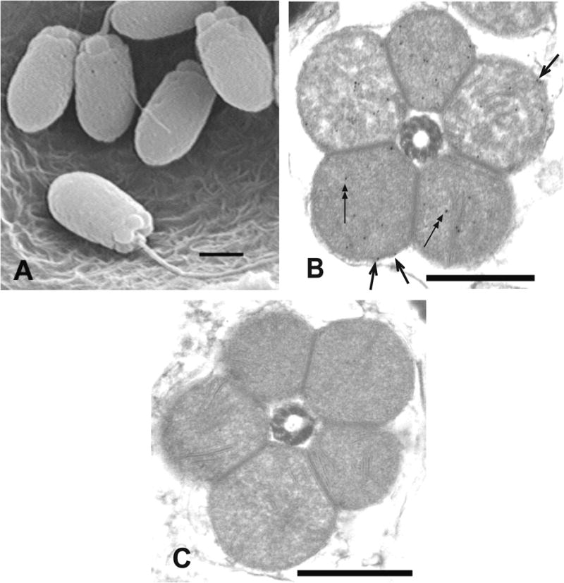
Figure 4. EM photomicrographs demonstrating unionoidean bivalve sperm morphology and the sub-cellular location of MCOX2 in sperm mitochondria.

(A) The general morphology of unionoidean bivalve spermatozoa (SEM of Plethobasus cyphyus sperm; scale bar = 1μm). (B) experimental IEM photomicrograph which displays immunogold labeling, using the MCOX2 primary antibody, in both the inner (two headed arrows) and outer (single headed arrows) mt membranes in a cross-section of the mitochondrial “ring” from Fusconaia subrotunda sperm, and (C) a negative control IEM photomicrograph from F. subrotunda sperm (both IEM scale bars = 0.5μm).
