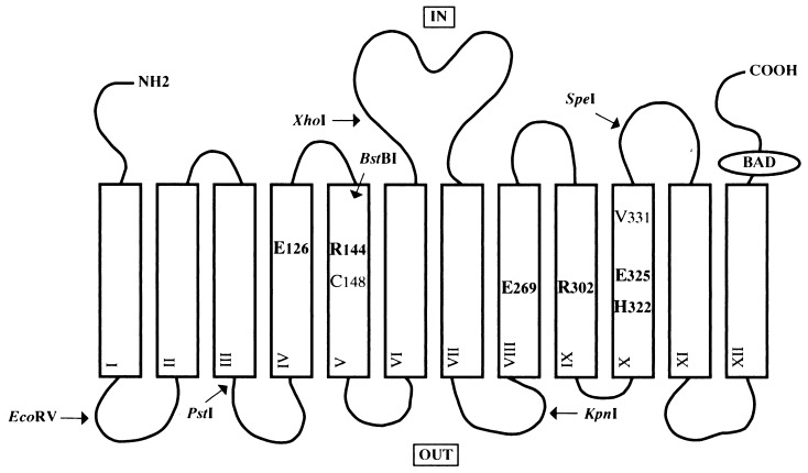
Figure 1.
Secondary structure model of lac permease. Putative transmembrane helices are shown in boxes. The positions of the six irreplaceable residues (Glu-126, Arg-144, Glu-269, Arg-302, His-322, and Glu-325), as well as Cys-148 and Val-331, are indicated. Also shown are the restriction endonuclease sites used for constructing the mutants and the biotin acceptor domain (BAD). The topology of helices IV and V have been modified as described in the text.

