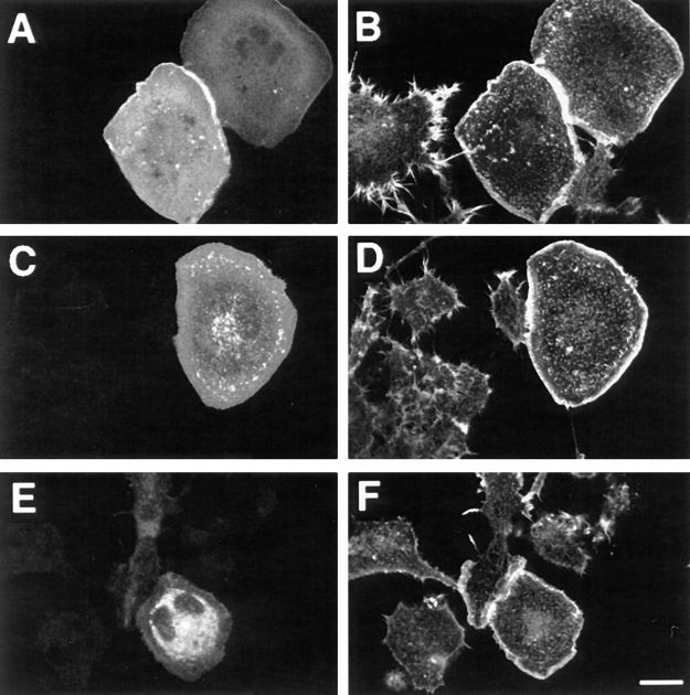
Figure 2.
Morphological characteristics of N1E-115 cells expressing Tiam1, Rac1, or Cdc42. Transiently transfected cells were grown on glass coverslips for two days, fixed, permeabilized, and stained with anti-Tiam1 antibodies (A) or anti-myc antibody 9E10 to detect V12Rac1 (C) and V12Cdc42 (E). TRITC-labeled phalloidin was used to detect F-actin (B, D, and F). Cells were viewed by confocal microscopy. The untransfected control cells are only visible in the panels stained for F-actin (B, D, and F). Bar, 25 μm.

