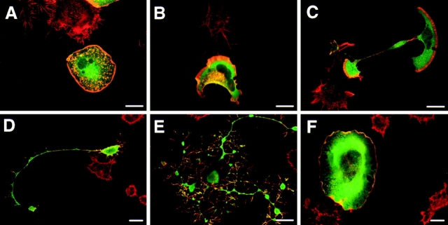
Figure 3.
Morphological transitions observed in Tiam1 cells grown on laminin. C1199 Tiam1–transfected cells were seeded onto laminin-coated coverslips in the presence of serum and allowed to adhere for various lengths of time before fixation and staining. Representative examples of each timepoint are given. (A) Spreading cell, 2 h after seeding. (B) Polarized cell 2 h after seeding, carrying a large lamellipodium at the leading edge. (C) Cell developing neurite-like outgrowths, 4 h after seeding. Note the presence of lamellipodia on both ends of the cell, moving in opposite directions. (D) Neurite-bearing cell after overnight growth on laminin. (E) Extreme branching induced by high expression of C1199 Tiam1 after overnight growth on laminin. (F) Some cells show extreme spreading and an apparent loss of contractility induced by high expression of C1199 Tiam1 after overnight growth on laminin. Tiam1 is shown in green, F-actin in red. Note that D and F represent a 400× magnification, whereas the other images were magnified 600×. The red cells represent untransfected controls. Bars, 25 μm.

