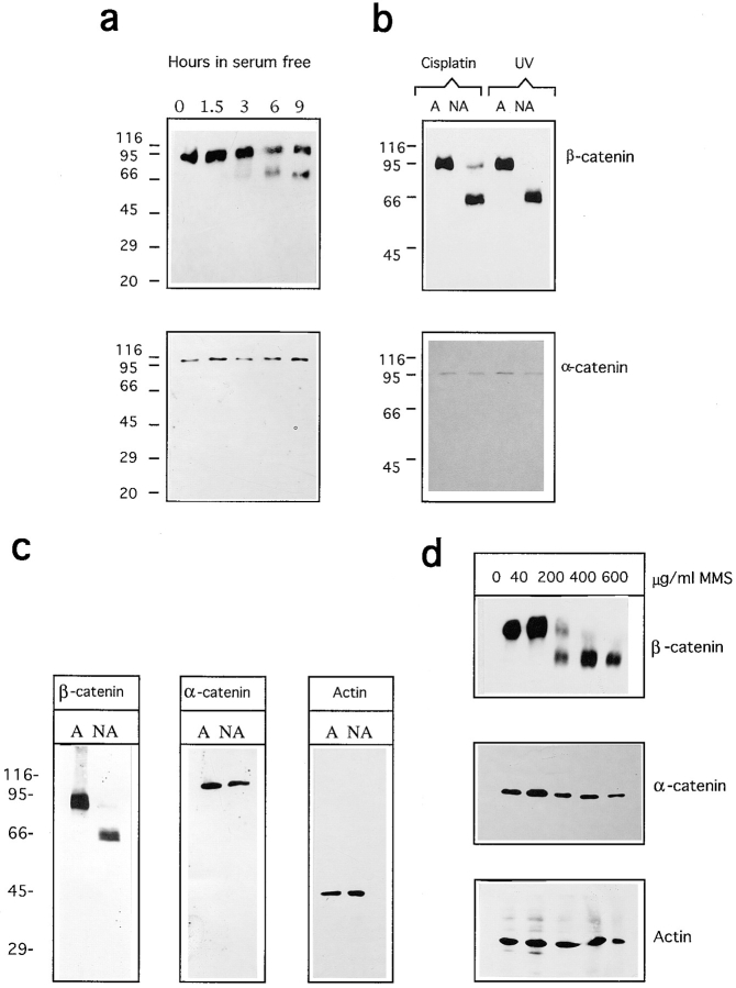
Figure 4.
Analysis of β-catenin cleavage during apoptosis in NIH3T3 and MDCK cells. (a) Growth-arrested NIH3T3 cells were incubated in serum-free medium for the indicated times. Lysates from both adherent viable cells and nonadherent apoptotic cells were combined and Western blot analysis was performed. (b) Density-arrested NIH3T3 cells were treated with 20 μg/ml of cisplatin or UV irradiated as described in Materials and Methods. After 24 h adherent (A) and nonadherent (NA) cells were processed separately and Western analysis was performed. (c) MDCK cells grown for 4 d in 10% FCS were incubated in serum-free medium for 12 h. Adherent (A) and nonadherent (NA) cells were processed separately and Western analysis was performed. (d) MDCK cells grown for 4 d in 10% FCS were treated for 4 h with the indicated amount of MMS. After 24 h, lysates from both adherent viable cells and nonadherent apoptotic cells were combined and Western blot analysis was performed.

