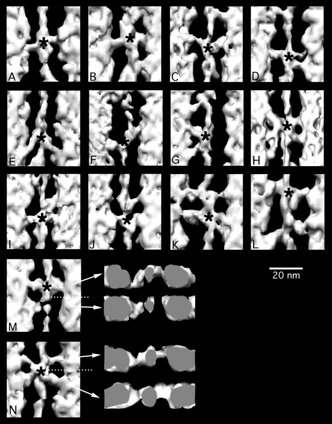
Figure 3.
Gallery of glycol-PNP crossbridges. Asterisks mark the axial positions of target zones. A, B, and D typify the most prevalent 90° axial attachment angle of target zone bridges. C, E, F, and G show the large axial angle variation that can be detected in target zone bridges. G also shows a partial mask motif and an irregular nontarget bridge. (H) Complete mask motif. (I) 90° target bridge pair and one mask bridge. (J–L) show target bridges and variably structured nontarget bridges. (M) Target and nontarget crossbridges with transverse view. The azimuth of the origin of the lower nontarget crossbridge in (M) and its approach to actin are the same as the upper target zone crossbridge, although the azimuths of their binding sites have rotated 81° and their predicted origins also differ. (N) Mask motif in horizontal and transverse view, showing that both bridge pairs originate from the thick filament and approach actin at similar azimuths. Transverse views look toward the Z-line. In longitudinal view, Z-line is at the bottom.

