Figure 5.
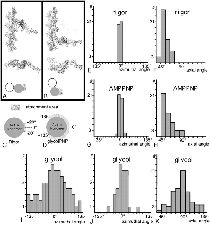
Comparison of the azimuthal and axial orientation of crossbridges in rigor, aqueous-PNP, and glycol-PNP. In A, the rigor acto-S1 coordinates are shown in longitudinal and in transverse view in their normal orientation. At the bottom, the shaded rectangle next to the filled circle shows the attachment area, the center of which we give the value of 0.0° as shown in C. (B) A nontarget zone crossbridge in a position often seen in the glycol-PNP reconstruction, two actin monomers below the lead target zone. The transverse view of B shows that the nontarget bridge attaches to a different part of the actin monomer than the rigor bridge in A. This azimuthal position would be assigned an angle of 70°. C illustrates the narrow range of azimuthal attachment areas on the actin monomer seen in rigor. D illustrates the broad range of azimuthal attachment areas when all glycol-PNP crossbridges are included. (E) Plot of the narrow distribution of the azimuthal contact of rigor lead bridges to actin. (F) Plot of the range of axial attachment angles seen in rigor. (G) Aqueous AMPPNP lead bridges also show a narrow azimuthal contact with actin. (H) Axial angles of aqueous AMPPNP lead bridges show a spread comparable to rigor but are slightly displaced toward 90°. (I) Plot of the distribution of azimuthal angles for all glycol-PNP bridges compared with (J) the distribution of target zone bridges only. (K) Plot of the range of axial attachment angles for glycol-PNP target zone bridges shows that 90° is the most frequent angle. Data for (E–H) were taken from reconstructions previously reported (Schmitz et al., 1996).

