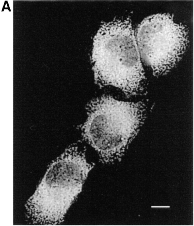
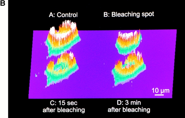
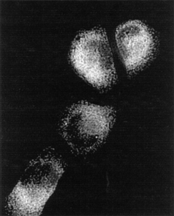
Figure 3.
Effects of the Ca2+-depletion protocol on the morphology and lumenal continuity of the ER as measured with erGFP. HeLa cells were transiently transfected with the erGFP construct and analyzed 2 d later. (A) ER morphology as revealed by erGFP fluorescence analyzed by confocal microscopy in live cells before (left) and 1 h after incubation in Ca2+-free, EGTA-containing KRB, and treatment with 30 μM tBuBHQ (right). (B) Photobleaching and recovery. After the depletion protocol was terminated, the image shown in A (control) was taken with the standard illumination protocol. A mask was then introduced in the exciting light path and the second image was taken (B; bleaching spot). The sample was then continuously illuminated with the highest laser power for 3 min before removing the mask and immediately taking the third image (C; 15 s after bleaching) with the standard settings. The last image (D; 3 min after bleaching) was collected under standard conditions, 3 min after the third. The cells were not illuminated during this recovery period. Bar, 9 μm.


