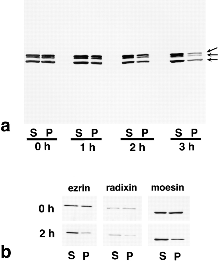
Figure 4.
Solubility of ERM proteins in LHF cells at 0, 1, 2, and 3 h after addition of FasL. LHF cells were homogenized in physiological saline and centrifuged to separate the soluble and insoluble fractions into the supernatant (S) and pellet (P), respectively. Equivalent amounts of supernatant and pellet were applied to SDS-PAGE and subsequently subjected to immunoblotting. To detect all members of ERM proteins, pAb TK89 was used (a). To detect ezrin, radixin, and moesin separately, mAb M11, mAb R21, and mAb M22 were used, respectively (b). ERM proteins translocated from the insoluble (P) to the soluble fraction (S) as apoptosis proceeded. Arrows in a indicate ezrin, radixin, and moesin, respectively, from the top.

