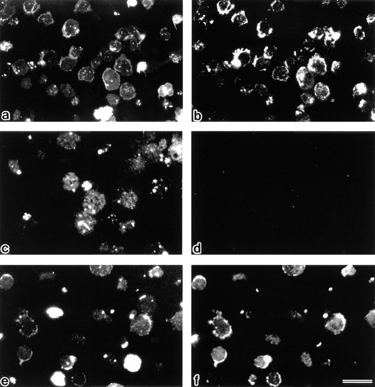
Figure 9.
Detection of ERM proteins on single-layered plasma membranes isolated on cover glasses. After vital labeling of plasma membranes proper with DiO, single-layered plasma membranes with the cytoplasmic surface freely exposed were prepared from nontreated (a and b), FasL–1 h–treated (c and d), and FasL/calyculin A–1 h–treated (e and f) LHF cells as described in Materials and Methods. These preparations were immunofluorescently labeled with anti-ERM pAb, TK89. Each single-layered plasma membrane was detected by DiO signal (a, c, and e). ERM signal was intense from plasma membranes of nontreated cells (b), whereas it was undetectable from those of FasL-treated cells (d). Calyculin A suppressed the FasL-induced depletion of ERM proteins from plasma membranes (f). Bar, 20 μm.

