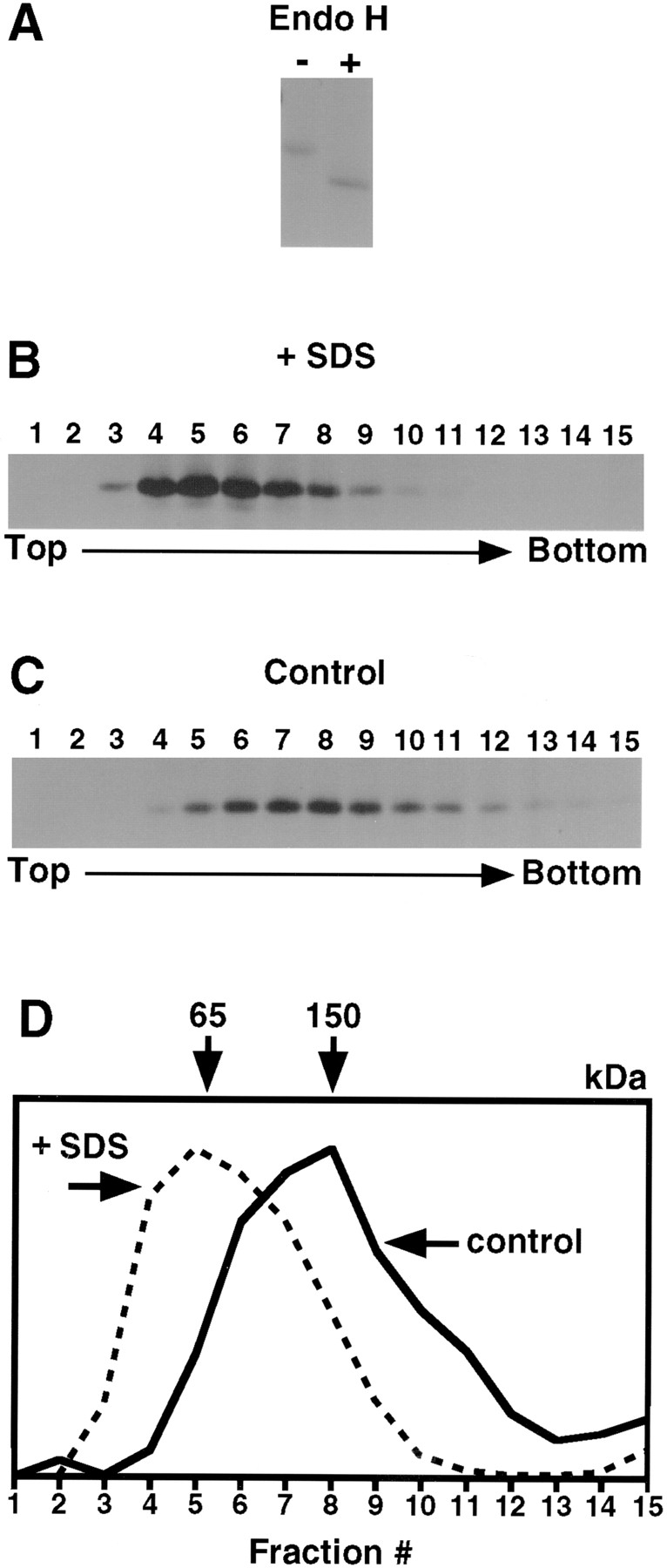
Figure 6.

The type II receptor forms homodimers in the ER. L6 cells were pulse-labeled with 35S-Express for 15 min, chilled, lysed with (B) or without (C) 0.5% SDS, and loaded on sucrose gradients with (B) or without (C) 0.5% SDS, as in Fig. 1. D is a graphic representation of the data as in Fig. 1. Endo H digestion of control material (A), labeled similarly, confirms that all labeled receptor is in the ER form (Endo H sensitive).
